|
Services - DiagnosticDiagnosis is the gateway to pinpointing disease and deciding the correct method of treatment. Imaging technologies are the most accurate and pervasive means of diagnosis available in our two areas of primary focus i.e. cancer and neuroscience. Imaging diagnostics are almost ubiquitous in medicine. The testing is done for problems as routinely mundane as broken bones to some of the most serious and severe forms of cancer such as glioblastoma mutiforme. We focus on this area of medicine because it compliments our current treatment facilities, it is widely used and acceptable as a technology and is a proven revenue generator with new applications always evolving. The diagnostic tools listed below are as multifaceted as they are acceptable to the medical community.
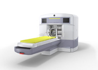
Computerized Tomography (CT) Computerized Tomography (CT)CT scans represent a major advance over the simple x-ray, which did not allow the doctor to visualize tissue at all. Instead of a flat, two-dimensional picture, CT scanners produce a series of successive images. These slices can be combined to create the illusion of depth and help to visualize if and where a problem exists. This is a mature marketplace yet still has significant potential. Magnetic Resonance Imaging (MRI)This technology takes advantage of the body's natural magnetic field, measuring change in the field's energy as patients are exposed to various radio frequencies. Unlike CT views, MRI's can be rendered in full 3-D because MRI machines can slice along three or more planes not just one. A computer can then compile the information to generate a sort of relief map of the site in question. This is another relatively mature market but there has been significant technological advancement with 3.0T MRI and High Field Open MRI, enhancing image quality and increasing patient throughput, thereby keeping this a very viable and continually growing marketplace. Positron Emission Tomography (PET)PET scans are an imaging technique that uses positively charged particles (radioactive activities). A PET scan provides a color-coded image of the body's function, metabolism and structure. This is probably the best and most expensive technique available today. It is a relatively new market with an extremely vast array of applications and advantages. This market technology is advancing rapidly with the advent of PET/CT, a combination of PET scans and CT scans which enhance the image quality and patient throughput. This market segment is growing at a swift rate Magnetic-Encephalography (MEG)This is an exciting, new technique that allows scientists to study the brain in real time. MEG directly measures nerve cell firing by detecting changes in tiny magnetic signals produced by active brain neurons. It is ideal for tracing the order and pattern in which brain regions are activated when performing certain tasks. Researchers now have the unprecedented ability to see what is going wrong with the brain and, perhaps, find cures for such diseases as epilepsy and many others. <<BackOverview - History - Gamma Knife - Partners - Services - Management - SEC Filings © Copyright 2005 U.S. NeuroSurgical |